
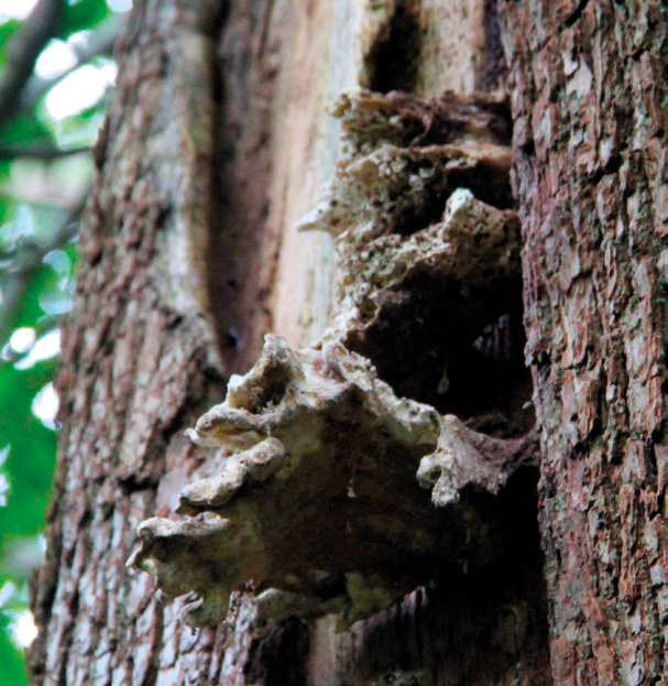
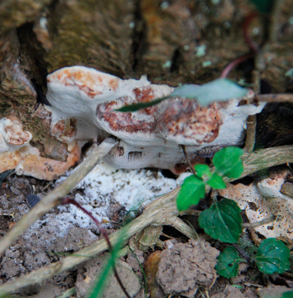
Brown rot present in one of the many standing monoliths at Clyne gardens.
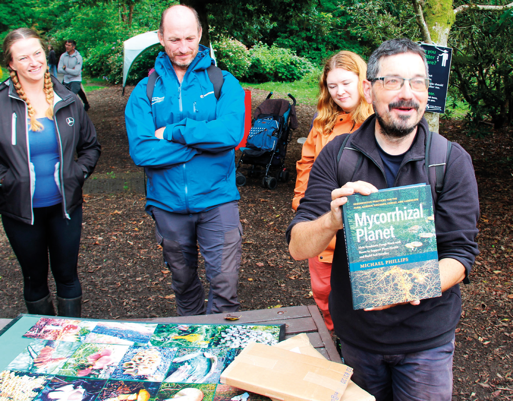
Teifion receiving his book – a thank you from the branch.
Mike Higgins, Wales Branch Chair
On 16th September, the Wales Branch held our fifth annual fungi walk at Clyne Gardens, Swansea. The event was again organised by Teifion Davies from Clyne Gardens and Steve Lucocq, Wales Branch Secretary.
Teifion is the Head Gardener at Clyne and is responsible for the 2,500 trees on site, a collection of 350-400 different species. Due to the inter-relationships between trees and fungi, it is likely that Clyne has more fungi than anywhere else in the city of Swansea.
Typically, fungi are identified by their appearance, but advances in DNA analysis are helping to identify species more accurately, and science and practical work are now improving understanding of the relationship between fungi and trees. Social media is also helping to communicate and share knowledge on these specialist subjects. However, the best way to gain experience is to visit sites where fungi can be seen, just like Clyne.
Clyne Gardens is now one of Wales’s 22 Sentinel Sites. These are public parks and gardens where the Animal and Plant Health Agency carries out plant health inspections twice a year. The parks feature thousands of different common and less well-known plants and trees, and the inspections note potential issues with damaging and regulated plant pests and diseases.
Teifion also talked about a large change in the management of trees at the site since he has worked there, with a focus now on the retention of standing decaying wood (monoliths aka snags) and laying larger stems to ground. The smaller material is also retained in the form of habitat piles. Throughout the site this could be seen, with excellent examples of monoliths and habitat piles as well as large, failed trees left in situ on the ground and being used by various other species such as invertebrates, epiphytes and fungi.
This year we had 12 attendees of varying backgrounds from arboricultural consultants to individuals with a general interest in fungi. We were again treated to an array of different fungi, some of which we have seen each year and some new ones.
As always, the event ended with Steve Lucocq presenting Teifion with a book as a gift of thanks. The book was fungi related as usual and will no doubt add to the knowledge Teifion has to share with the group next year.
All images by Mike Higgins
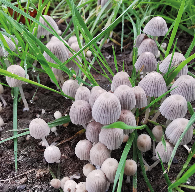
1. Trooping crumble cap (Coprinellus disseminatus)
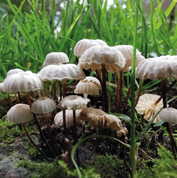
2. Collared parachute fungi (Marasmius rotula)
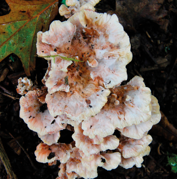
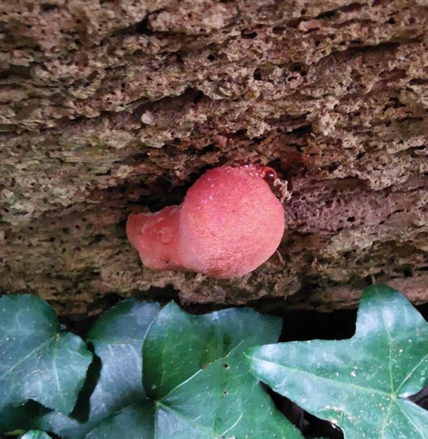
3a. Blushing rosette (Abortiporus biennis)
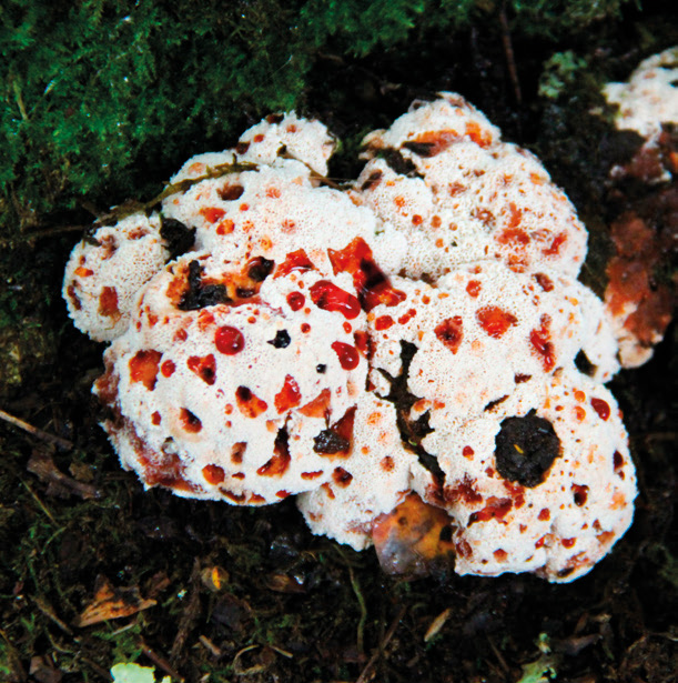
3b. Blushing rosette (Abortiporus biennis)
- Trooping crumble cap (Coprinellus disseminatus) growing on the ground next to the Sentinel noticeboard.
- Collared parachute fungi (Marasmius rotula) growing on the decaying root of an ash tree. The group was able to follow the path of the root based on the fruit bodies. The root was likely damaged by mowers as it breached the surface, allowing the fungi to colonise.
- Blushing rosette (Abortiporus biennis) on red oak with typical form (a) and an abortive form with stunning guttation (b). We have been lucky to see this fungus every year the walk has taken place. It was also found on the stump of a katsura tree at another location on the site.
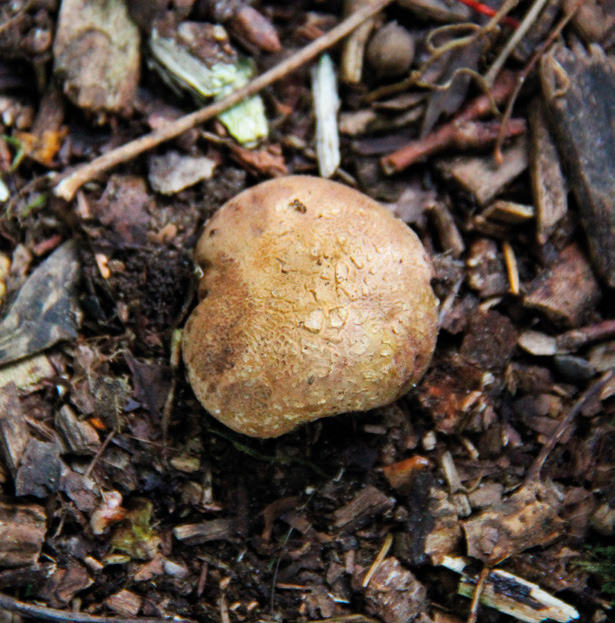
4. Common earthball (Scleroderma citrinum)
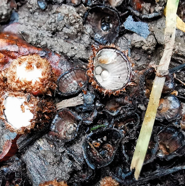
5. Fluted bird’s nest (Cyathus striatus)
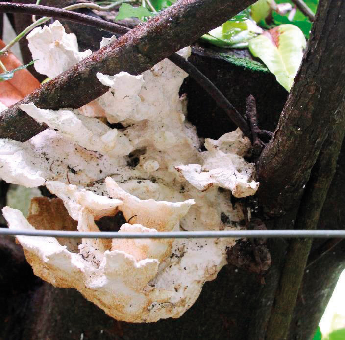
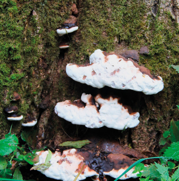
6a. Chicken of the woods (Laetiporus sulphureus)
6b. Chicken of the woods (Laetiporus sulphureus)
- Common earthball (Scleroderma citrinum).
- Fluted bird’s nest (Cyathus striatus). This specimen raised the issue of how different fungi spread their spores. C. striatus uses raindrops and other fascinating means by which to reproduce. The evolution of the fruit body is such that it is the correct size that when a raindrop hits the centre, it forces the peridioles (‘egg’) out of the ‘nest’ structure. The egg has a funicular (microscopic hyphal cord) which attaches onto adjacent plants rather than hitting the ground. Once attached, the ‘egg’ matures until the spores are finally released. However, this is rarely a means of reproduction as two mycelia must make contact, and this colliding/mating on adjacent plants is typically the most successful method.
- Chicken of the woods (Laetiporus sulphureus). A mature, pale form unusually spotted growing on cherry laurel (a) and a remnant specimen at a height of 5 metres on an oak (b). The specimen on the cherry laurel is likely to have established due to exposed heartwood following a previous topping of the host. The group discussed colonisation strategies and biodiversity potential from fungi and tree relationships. Heartwood decay tends to be a natural phenomenon which in turn produces beneficial habitat for invertebrates and birds etc. This type of habitat was observed throughout the site on living trees, as well as on standing monoliths/snags and failed decaying wood on the ground.
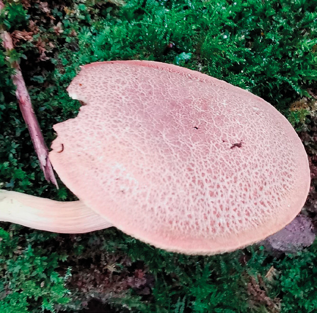
7a. Suede bolete (Xerocomus subtomentosus)
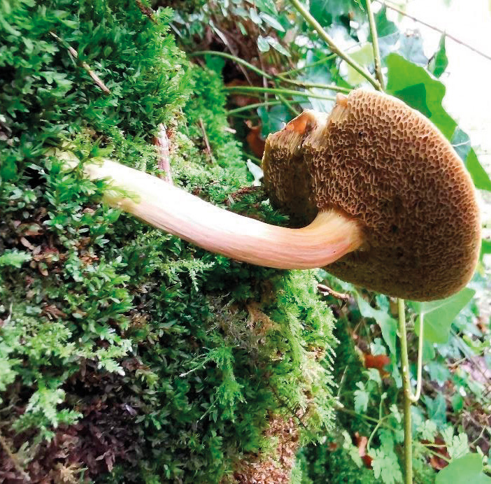
7b. Suede bolete (Xerocomus subtomentosus)
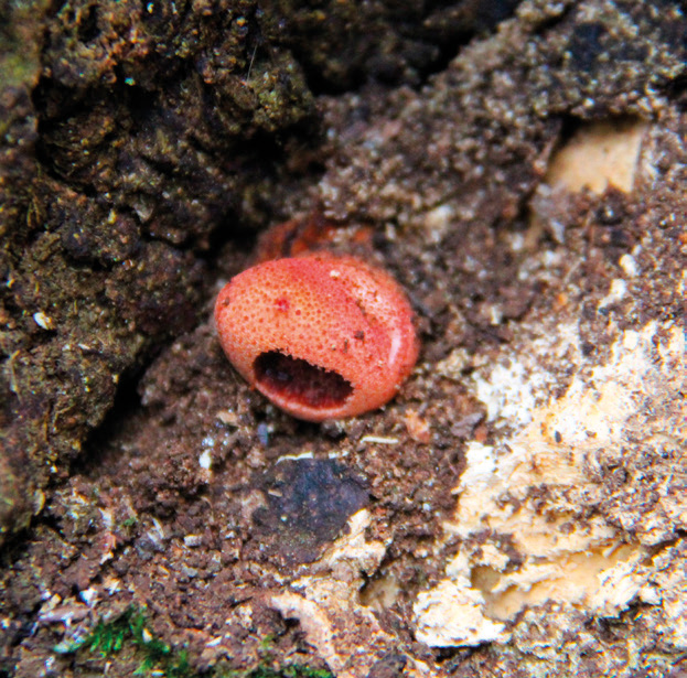
8a. Beefsteak fungus (Fistulina hepatica)
8b. Beefsteak fungus (Fistulina hepatica)
- Suede bolete (Xerocomus subtomentosus). This particular specimen was growing on the bank at the edge of a wooded area in the gardens. The unusual location made it a bit easier for the group to see the yellow/brown tubes and pores on the underside.
- Beefsteak fungus (Fistulina hepatica). A small emerging specimen on a failed tree (a) and another on the underside of another retained failed trunk elsewhere on site (b).
9a. Ganoderma sp.
9b. Ganoderma sp.
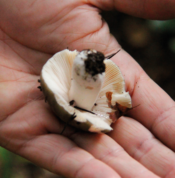
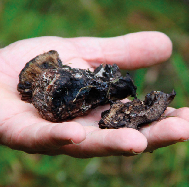
10. Brittlegill (Russula sp.)
11. Yew duster fungus (Amylostereum laevigatum)
- Ganoderma sp. on ash (a) and on a fallen stem (b). The Ganoderma instigated a discussion between the arboriculturists in the group on the difficulties of distinguishing G. applanatum from G. australe. One of the main ways to distinguish G. applanatum is the presence of galls on the underside caused by a platypezid fly (Agathomyia wankowiczii). The galls can be felt underneath as small, quite sharp protuberances. The group noted that the presence of galls is not common in West Wales. As one of the locations included a bracket on a failed stem, the discussion also turned to geotropism, and the process by which a bracket will form parallel to the ground, which informs the observer that the bracket in question formed after the tree failed.
- Brittlegill (Russula sp.) is a common woodland mycorrhizal fungus. This specimen showed it is a tasty snack for slugs. It is pictured in Teifion’s hand to show the group the reason for the name – numerous intricate, delicate gills make up the underside of the fruit body.
- Yew duster fungus (Amylostereum laevigatum). This was a special find as it is the first time it has been recorded at Clyne gardens. The species is a quite unassuming crust fungus that was growing on the underside of a single yew branch about 1.5 metres from the ground. The weather was quite wet and overcast so it was quite difficult to spot, but thankfully Teifion knew where to look.
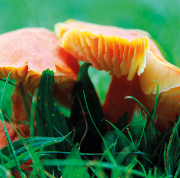
12. Fibrous waxcap (Hygrocybe intermedia)
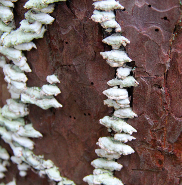
13. Purplepore bracket (Trichaptum abietinum)
14. Blackening russula (Russula nigricans)
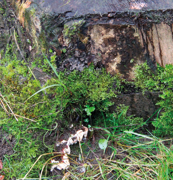
15a. Root rot fungus (Heterobasidion annosum)
- Fibrous waxcap (Hygrocybe intermedia). This was the last find before lunch and is one of the non-mycorrhizal fungi found on the day. This is more common in Wales than most of Britain.
- Purplepore bracket (Trichaptum abietinum). This is another saprotrophic species to benefit from the more biodiverse management of the trees in Clyne, being found in abundance on a decaying pine monolith. The monolith was also showing signs of insect activity with holes and galleries present.
- Blackening russula (Russula nigricans). A mature fruit body found on site and although not a perfect specimen, it was noted as it is often associated with a parasitic fungus (Asterophora parasitica), although we were not lucky enough to see it this time, so something to look for next year.
- Root rot fungus (Heterobasidion annosum). This specimen (a) is a little confusing at first sight as it appears to be growing at the base of a hazel coppice. However, it is likely that it is actually growing on an historical stump or root remnant of a conifer submerged below the hazel. The smaller specimen pictured (b) was growing from the stump of a Nothofagus and the fungi was also spotted during the day on the base of a Cupressus macrocarpa stump.
15b. Root rot fungus (Heterobasidion annosum)
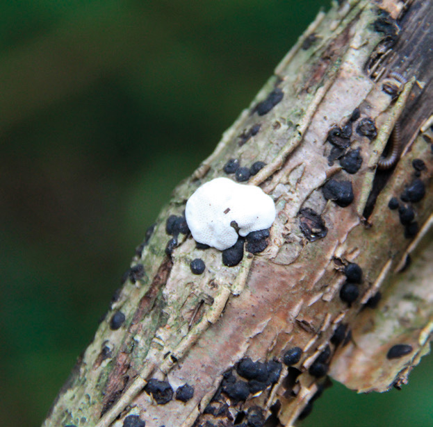
16. Hazel woodwart (Hypoxylon fuscum)
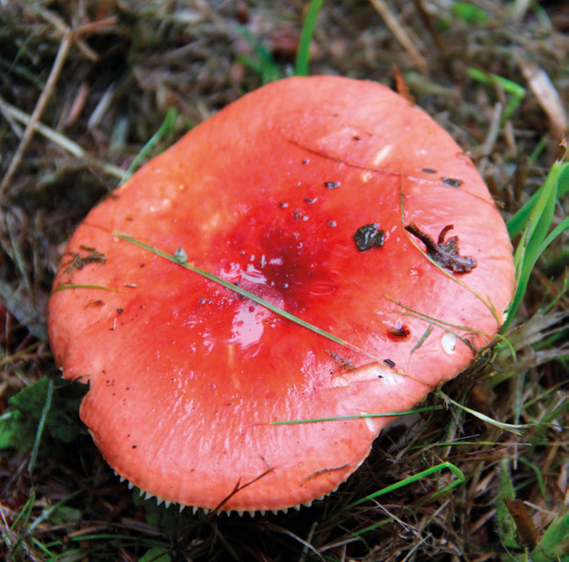
17a. The sickener (Russula emetica)
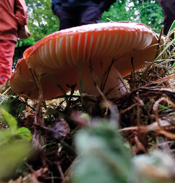
17b. The sickener (Russula emetica)
- Hazel woodwart (Hypoxylon fuscum). This saprotrophic fungus is the small black lumps in the picture on the decaying branch of a hazel, making identification more straightforward if you know the tree it is growing on.
- The sickener (Russula emetica). This mycorrhizal specimen was like a beacon in the undergrowth. Teifion explained that the name is an accurate description: eating it will cause sickness.
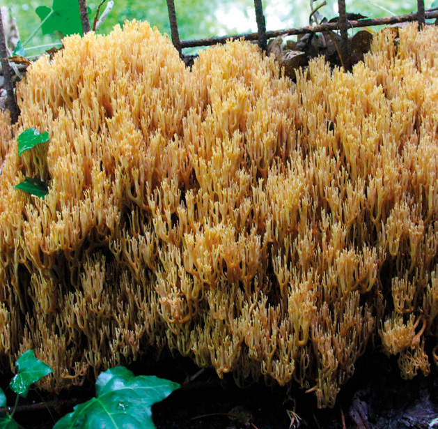
18. Upright coral fungus (Ramaria stricta)
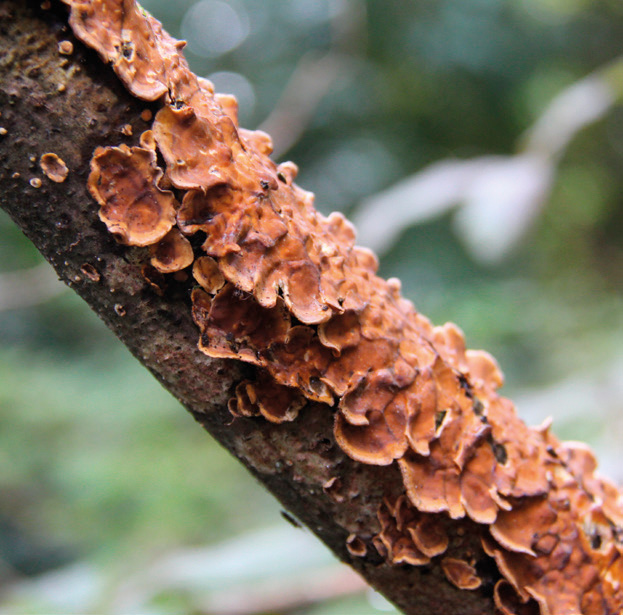
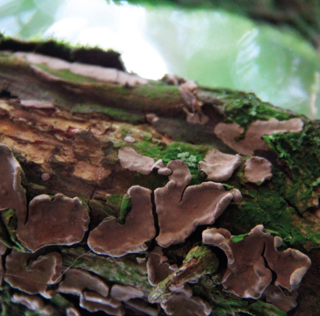
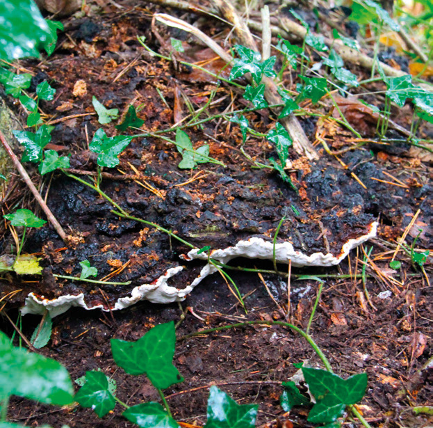
19. Hairy curtain crust (Stereum hirsutum)
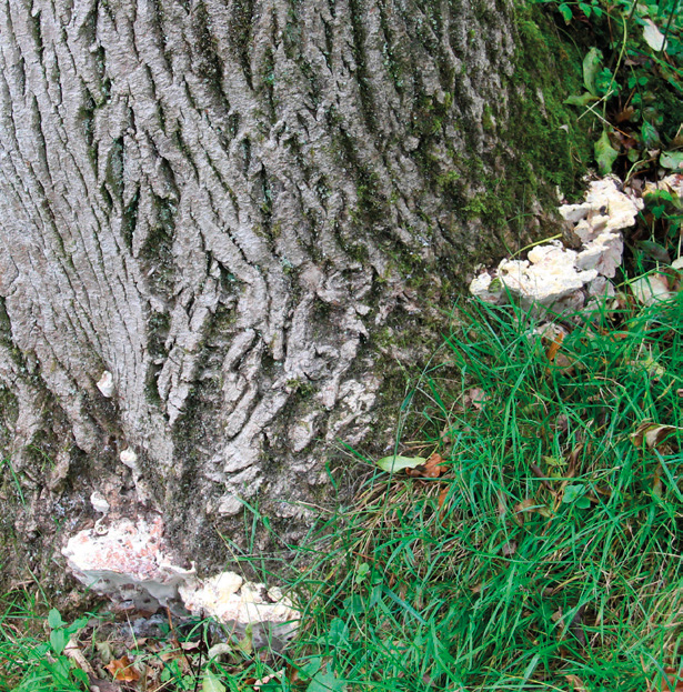
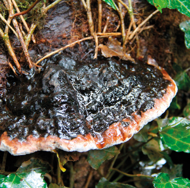
20a. Giant ash bracket (Perenniporia fraxinea)
20b. Giant ash bracket (Perenniporia fraxinea)
- Upright coral fungus (Ramaria stricta). This was the highlight of the day and Teifion saved it for last. This is an unusual fungus in that it can be saprotrophic and mycorrhizal. This emergence was growing on the top of a woodland bank next to an old fence and spread out along the fence in scattered pockets for about 2 metres.
- Hairy curtain crust (Stereum hirsutum). Found growing on the top of a decaying oak branch lying on the ground, it was predominantly in the resupinate crust form, although this species is often found in stacked tiers. The common name comes from the wavy edge that resemble curtains and the hirsutum comes from the downy covering on the younger fruiting bodies. This could be seen on a few of the smaller ones, although most of those on the branch had lost their downy appearance.
- Giant ash bracket (Perenniporia fraxinea). Found on a mature ash on the site with fruiting bodies present around the entire circumference of the base of the tree (a). One of the specimens was also sporulating (b).
Note: The white fungus in the image is the emerging form of a different species but it was too small to positively identify.
This article was taken from Issue 203 Winter 2023 of the ARB Magazine, which is available to view free to members by simply logging in to the website and viewing your profile area.